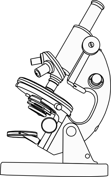
42 microscope diagram without labels
en.wikipedia.org › wiki › Electron_microscopeElectron microscope - Wikipedia An electron microscope is a microscope that uses a beam of accelerated electrons as a source of illumination. As the wavelength of an electron can be up to 100,000 times shorter than that of visible light photons, electron microscopes have a higher resolving power than light microscopes and can reveal the structure of smaller objects. Microscope Diagram Labeled, Unlabeled and Blank | Parts of a Microscope Description Worksheet identifying the parts of the compound light microscope. Answer key: 1. Body tube 2. Revolving nosepiece 3. Low power objective 4. Medium power objective 5. High power objective 6. Stage clips 7. Diaphragm 8. Light source 9. Eyepiece 10. Arm 11. Stage 12. Coarse adjustment knob 13. Fine adjustment knob 14. Base S
› en › microscopeFluorescence Resonance Energy Transfer (FRET) Microscopy Presented in Figure 3 is a Jablonski diagram illustrating the coupled transitions involved between the donor emission and acceptor absorbance in fluorescence resonance energy transfer. Absorption and emission transitions are represented by straight vertical arrows (green and red, respectively), while vibrational relaxation is indicated by wavy ...

Microscope diagram without labels
PDF Parts of a Microscope Printables - Homeschool Creations Label the parts of the microscope. You can use the word bank below to fill in the blanks or cut and paste the words at the bottom. ... without needing to move the microscope ? the head •What is the magnification level on the eyepiece of a microscope?10x (see objective Parts of the Microscope with Labeling (also Free Printouts) Parts of the Microscope with Labeling (also Free Printouts) A microscope is one of the invaluable tools in the laboratory setting. It is used to observe things that cannot be seen by the naked eye. Table of Contents 1. Eyepiece 2. Body tube/Head 3. Turret/Nose piece 4. Objective lenses 5. Knobs (fine and coarse) 6. Stage and stage clips 7. Aperture › topics › medicine-andConfocal Microscopy - an overview | ScienceDirect Topics A standards document, which describes confocal microscopy and its influence quantities, has recently completed an ISO ballot as a final draft international standard (ISO FDIS 25178-607, 2018). A schematic diagram of a typical confocal microscope is shown in Fig. 15.1 (ASME B46-2009, 2010; Weller et al., 2012). Most examples of this method rely ...
Microscope diagram without labels. Microscope Labeling - The Biology Corner 1) Start with scanning (the shortest objective) and only use the COARSE knob . Once it is focused… 2) Switch to low power (medium) and only use the COARSE knob . You may need to recenter your slide. Once it is focused.. 3) Switch to high power (long objective). filtration diagram without label - shiraneh-esf.ir Label the microscope - Science Learning Hub Maximum Operating Pressure 50 psi. On the diagram below label the major parts of the kidney. It is a diagram including all forces acting on a given object without the other object in the system. While preparing tea, a filter or a sieve is used to separate tea leaves from the water. Labeling the Parts of the Microscope | Microscope World Resources Labeling the Parts of the Microscope This activity has been designed for use in homes and schools. Each microscope layout (both blank and the version with answers) are available as PDF downloads. You can view a more in-depth review of each part of the microscope here. Download the Label the Parts of the Microscope PDF printable version here. Microscope Poster - Diagram with Labels | Teach Starter A poster containing a diagram with labels showing the key parts of a microscope. In Science it is important that students know how to use a variety of tools when conducting scientific experiments and inquiry. This poster focuses on the microscope and highlights its key parts. There are two print options available for this poster:
Fluorescence - Wikipedia Fluorescence is the emission of light by a substance that has absorbed light or other electromagnetic radiation.It is a form of luminescence.In most cases, the emitted light has a longer wavelength, and therefore a lower photon energy, than the absorbed radiation. A perceptible example of fluorescence occurs when the absorbed radiation is in the ultraviolet … Microscope, Microscope Parts, Labeled Diagram, and Functions The Iris Diaphragm is located above the condenser lens and below the microscope stage. The different sized holes in the diaphragm helps to vary the size of the cone and intensity of light that is projected upward into the slide. However, there is no set rule regarding which setting to use for a particular power. Parts of Stereo Microscope (Dissecting microscope) – labeled diagram ... Labeled part diagram of a stereo microscope ... (based on color bands and their respective labels), the objectives of a dissecting microscope are located in a cylindrical cone and, therefore, are not directly seen. ... but large enough to be seen or handled without the aid of a high power compound microscope. Thus, stereo microscopes have a ... › books › NBK26880Looking at the Structure of Cells in the Microscope A special sample holder is used to keep this hydrated specimen at -160°C in the vacuum of the microscope, where it can be viewed directly without fixation, staining, or drying. Unlike negative staining, in which what is seen is the envelope of stain exclusion around the particle, hydrated cryoelectron microscopy produces an image from the ...
Answered: Learning through Art: Water Molecules… | bartleby Science Biology QA Library Learning through Art: Water Molecules and Hydrogen Bonding Part A Drag the labels to their appropriate locations in this diagram. • First use the labels of Group 1 to identify the atoms and charges. . Then use the labels of Group 2 to identify the bonds. Labels can be used once, more than once, or not at all. ionic bond O hydrogen bond nonpolar … pages.zeiss.com › rs › 896-XMS-794Principles of Fluorescence and Fluorescence Microscopy fies the principle of the fluorescence microscope — without the light-filtering abilities of the purple glass window and the glass of white wine, Stokes would not have been able to observe any fluorescence at all. Using Stokes’ observation and the green fluorescent protein (GFP) as examples, this article will explain Confocal Microscopy - an overview | ScienceDirect Topics A confocal microscope was invented in 1951 by Marvin Minsky, a postdoctoral fellow at Harvard University studying neural networks in living brain (Minsky, 1988).In 1957, Minsky patented the concept of confocal imaging, the illumination and detection of a single diffraction-limited spot in a specimen (Fig. 1A).In the transmission configuration, the condenser is replaced with a second … Simple Microscope - Parts, Functions, Diagram and Labelling A simple microscope is a device that only has one lens for magnification. It functions the same way as the magnifying glass. Although it is simple in terms of design and function, it is useful I various fields including medicine, jewelry and watchmaking, and agriculture, to name a few. References
Under the Microscope In Your Food: Milk and Bacteria
Label the microscope — Science Learning Hub In this interactive, you can label the different parts of a microscope. Use this with the Microscope parts activity to help students identify and label the main parts of a microscope and then describe their functions. Drag and drop the text labels onto the microscope diagram.

Minds Eye
7th grade Science - Microscope Diagram | Quizlet The Parts of a Microscope. 12 terms. totobear PLUS. Sets found in the same folder. Science Key terms 7th grade. 13 terms. palocastillo. 7th Grade Earth Science. 9 terms. EliseC17. 7thGrade Review - Cells/Biology. 26 terms. SolizScience TEACHER. 7th grade Science, Cell theory. 8 terms. Super1412. Other sets by this creator.
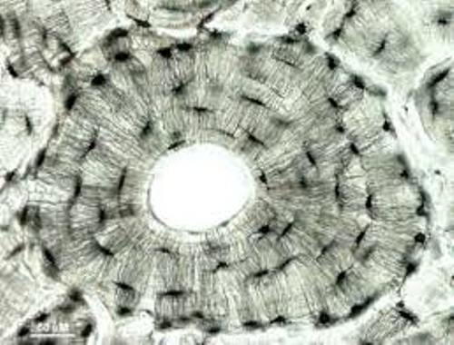
10 Facts about Bone Cells | Fact File
Labelled Diagram of Compound Microscope - Biology Discussion The below mentioned article provides a labelled diagram of compound microscope. Part # 1. The Stand: The stand is made up of a heavy foot which carries a curved inclinable limb or arm bearing the body tube. The foot is generally horse shoe-shaped structure (Fig. 2) which rests on table top or any other surface on which the microscope in kept.

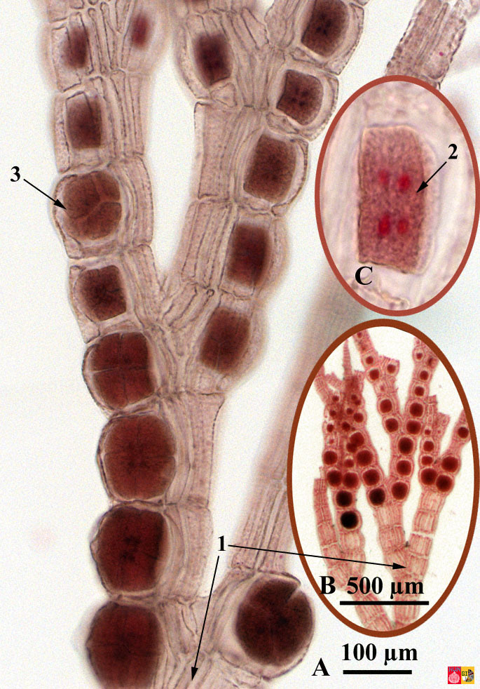
Transport in Plants
PDF Label parts of the Microscope: Answers Label parts of the Microscope: Answers Coarse Focus Fine Focus Eyepiece Arm Rack Stop Stage Clip . Created Date: 20150715115425Z ...
Microscope Clip Art at Clker.com - vector clip art online, royalty free & public domain
Compound Microscope Parts - Labeled Diagram and their Functions - Rs ... The eyepiece (or ocular lens) is the lens part at the top of a microscope that the viewer looks through. The standard eyepiece has a magnification of 10x. You may exchange with an optional eyepiece ranging from 5x - 30x. [In this figure] The structure inside an eyepiece. The current design of the eyepiece is no longer a single convex lens.

Compound Microscope Clip Art at Clker.com - vector clip art online, royalty free & public domain
Compound Microscope Parts, Functions, and Labeled Diagram Compound Microscope Definitions for Labels. Eyepiece (ocular lens) with or without Pointer: The part that is looked through at the top of the compound microscope. Eyepieces typically have a magnification between 5x & 30x. Monocular or Binocular Head: Structural support that holds & connects the eyepieces to the objective lenses.

A typical animal cell (as seen in an electron microscope) Medical Ima…
Microscope Label Interactive Worksheets & Teaching Resources | TpT 12. $1.89. PDF. Students will complete a timeline of the history of the microscope, label a diagram, and create a pocket foldable with terms and definition cards. The timeline can be completed according to the teacher's directions or like the answer key example. Optional cut & paste images and a QR code are a.
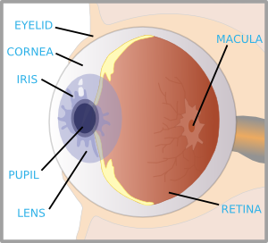
Eye With Labels Clip Art at Clker.com - vector clip art online, royalty free & public domain
Microscope labeled diagram - SlideShare Microscope labeled diagram 1. The Microscope Image courtesy of: Microscopehelp.com Basic rules to using the microscope 1. You should always carry a microscope with two hands, one on the arm and the other under the base. 2. You should always start on the lowest power objective lens and should always leave the microscope on the low power lens ...
Search in gallery
Multiphoton Microscopy | Nikon’s MicroscopyU Two-photon excitation microscopy (also referred to as non-linear, multiphoton, or two-photon laser scanning microscopy) is an alternative to confocal and deconvolution microscopy that provides distinct advantages for three-dimensional imaging.In particular, two-photon excitation excels at imaging of living cells, especially within intact tissues such as brain slices, embryos, whole …
Post a Comment for "42 microscope diagram without labels"